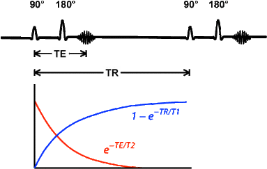
TE = time of echo = time from centre of RF pulse to centre of echo
TR = time of repetition = time between repeated pulses for repeating echo sequences

Echo magnitude = $S = K \; |H| \; e^{-\textrm{TE} / \textrm{T2}} \; (1 - e^{-\textrm{TR} / \textrm{T1}})$
Above, $(1 - e^{-\textrm{TR} / \textrm{T1}})$ is the magnitude of the $M_z$ just before the 90-degree flip (i.e. how much $M_z$ has recovered from the previous echo). That is the magnitude of the initial $M_{xy}$ after the flip. Of that initial $M_{xy}$, the fraction $e^{-\textrm{TE} / \textrm{T2}}$ is how much is then left at time TE after the flip. $|H|$ is the number of protons (since signal strength is proportional to the number of protons) and $K$ is some constant.
How does variance in TE affect the signal?
Suppose tissue A has short T2 and tissue B has a bit longer T2.
The $e^{-t / \textrm{T2}}$ decay is different for each.
With short TE, the echo occurs soon and the two different amplitudes cannot be distinguished.
With longer TE, the two curves (T2$_\textrm{A}$ and T2$_\textrm{B}$) will separate more and the echo will have more easily distinguished amplitudes.
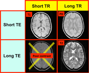
Longer TE gives more weight to T2 differences, so this produces a "T2 weighted" signal.
e.g. TE = 90ms, TR = 4000ms
Images can be distinguished with bright CSF.
Note, however, that the curves will come together again after a while. Maximum separation occurs at
$\begin{array}{rl} {d \over dt} \; (e^{-t/\textrm{T2}_A} - e^{-t/\textrm{T2}_B}) & = 0 \\ {e^{-t / \textrm{T2}_A} \over \textrm{T2}_A} - {e^{-t / \textrm{T2}_B} \over \textrm{T2}_B} & = 0 \\ t & = {\textrm{T2}_A \; \textrm{T2}_B \; \ln {\textrm{T2}_B \over \textrm{T2}_A} \over \textrm{T2}_B - \textrm{T2}_A} \\ \end{array}$e.g. liver T2$_A$ = 40ms, fat T2$_B$ = 70ms, maximum separation at TE = 52ms.
How does variance in TR affect the signal?
Tissue A with short T1 and tissue B with a bit longer T1
With long TR, both tissues have had time to recover their alignment with B0 after the 90-degree pulse, so they cannot be differentiated.
With shorter TR, tissue A (short T1) will recover move $B_0$ alignment than tissue B (long T1).
Shorter TR gives more weight to T1 differences, so this produces a "T1 weighted" signal.
e.g. TE = 14ms, TR = 500ms
Images can be distinguished with dark CSF.
So "T1 weighting" and "T2 weighting" do not yield images with pixel values proportional to some combination of the T1 values or T2 values.
