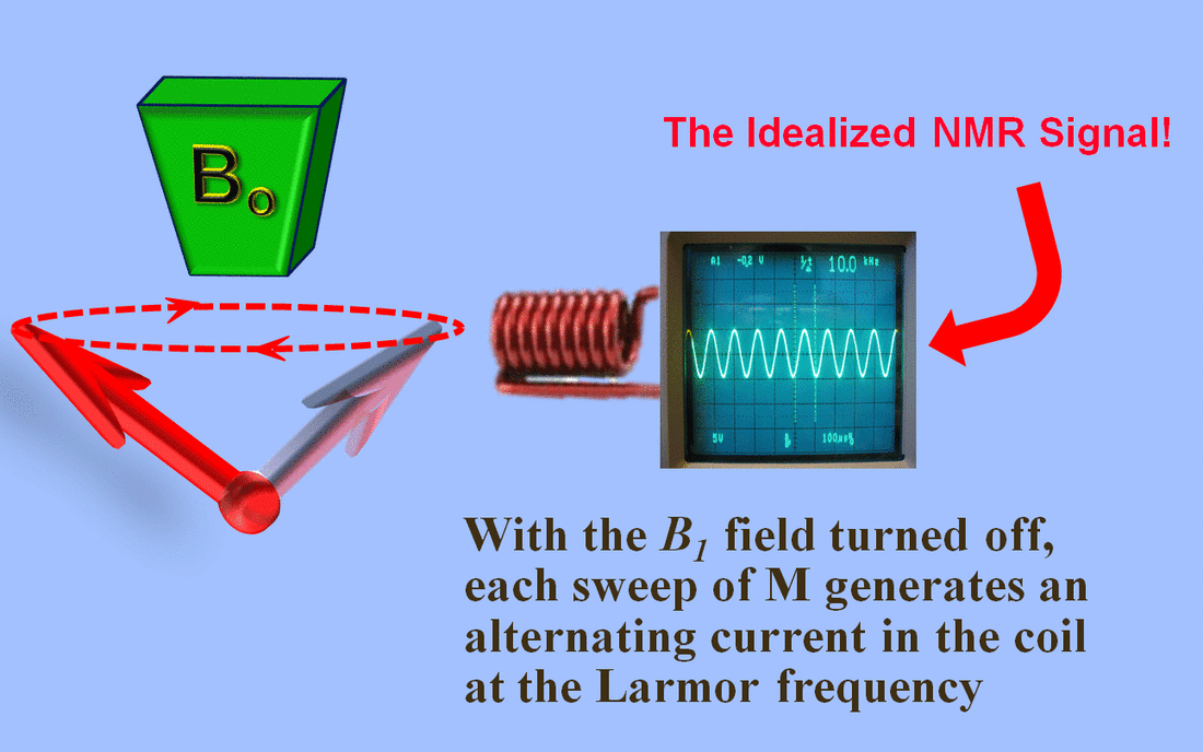
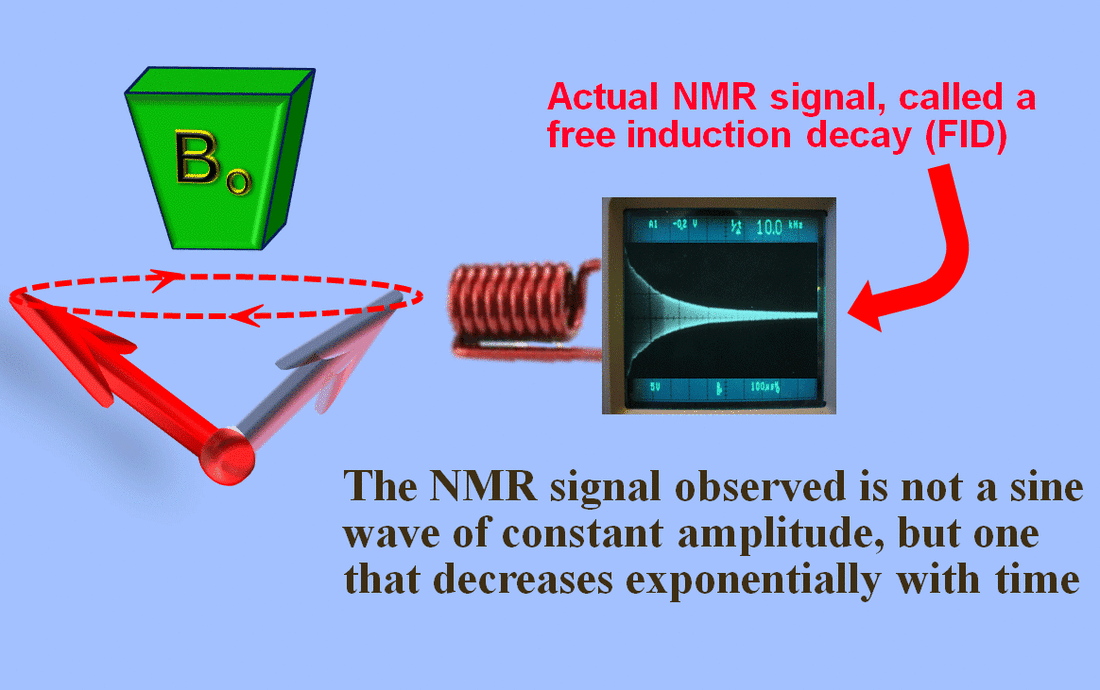
When $B_1$ is turned off, atoms interact with environment and lose energy. Upon energy loss, their magnetic moment re-aligns with $B_0$. This is called relaxation.
The thermal energy loss is tiny in the human body, so no harm comes to the subject.
We can split $M$ into two components: $M_z$ which is aligned with the $B_0$ axis, or the $z$ axis as it is also labelled; and $M_{xy}$ which is perpendicular to $M_z$:
$M = M_z + M_{xy}$
After $B_1$ is turned off, the precession continues with the same (Larmor) frequency and same phase, but the net magnetic moment, $M$, "tilts up" to align with $B_0$.
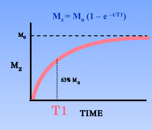
- The magnitude of $M_z$ increases as $|M_z| = |M_0|\ (1 - e^{-{t \over \textrm{T1}}})$, which we've seen before.
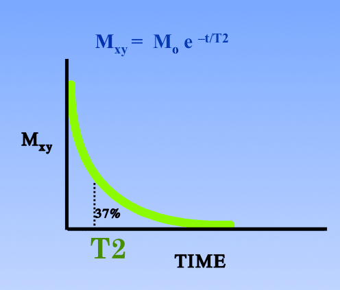
- The magnitude of $M_{xy}$ decreases as $|M_{xy}| = |M_0|\ e^{-{t \over \textrm{T2}}}$, which is new.
As before, T1 is the time that $|M_z|$ takes to reach about 63% of $|M_0|$.
And T2 is the time that $|M_{xy}|$ takes to reach about 37% of $|M_0|$.
This means that T2 is always less than T1:
T1 is in [0.1, 3.0]. T1 is the time constant for the net magnetic moment, $M_z$, in the $B_0$ direction.
T2 relaxation occurs from interactions with spins of close particles and local changes in the magnetic field, both of which cause a proton to fall out of phase with the rest of the in-phase protons.
T1 and T2 depend on molecule containing the $^1H$. Small fast molecules have long T1 and T2. Slow molecules get shorter T2 and longer T1:
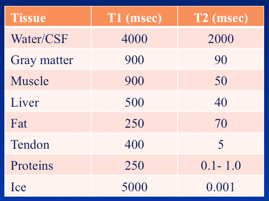
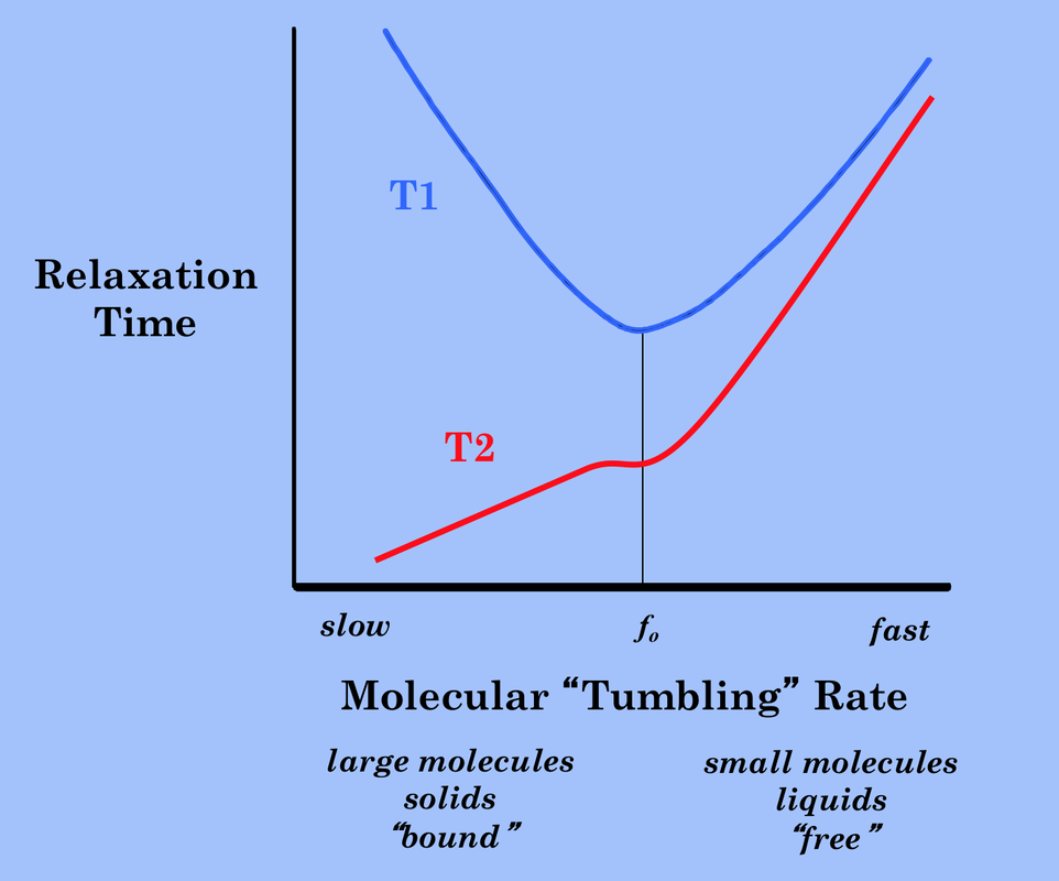
Longitudinal relaxation (T1) induces transverse relatation (T2) because the net spin, as it lines up with $B_0$, gets a smaller component in the transverse direction. But additional factors contribute to transverse relaxation, so it's usually faster (but never slower).
