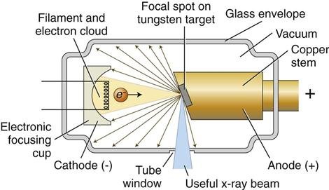
Heated cathode
Generates electrons
Voltage field
Accelerates electrons toward anode
Rotating anode
Can rotate for cooling; can also have water cooling.
Two types of xray interaction at anode:
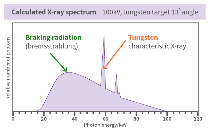
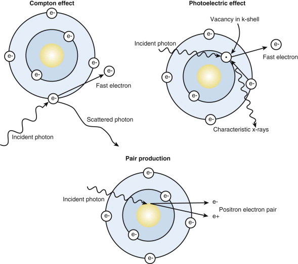
- characteristic: inner shell electon is dislodged; outer electron drops and emits photon with difference in energies; characteristic lines in spectrum which depends upon anode material
- Bremsstrahlung: "braking radiation"; given when electrons are scattered in electric field near heavy atoms. Decrease in electron energy near nucleus results in xray radiation at all energy levels
However: In mammography, low energy xrays are needed because fat, calcium, and soft tissues are best detected around 4 keV, so a molybdenum anode can be used instead of the usual tungsten anode; molybdenum has lower-energy characteristic xrays, which give better contrast for fat, calcium, and other soft tissue.
Filter Window
Can be thin metal or glass to remove low-energy xrays, which would otherwise be completely absorbed by patient.
However: In mammography, low energy xrays need to be preserved, so another material (e.g. beryllium) is used which does not have much attenuation at low energies.
Receiver
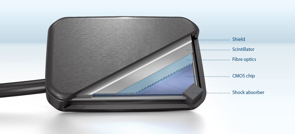
Can be:
- film (not used much now)
- scintillator: xrays become visible light, which are, in turn, sensed by a CCD array
- purely digital receivers (much better dynamic range)
