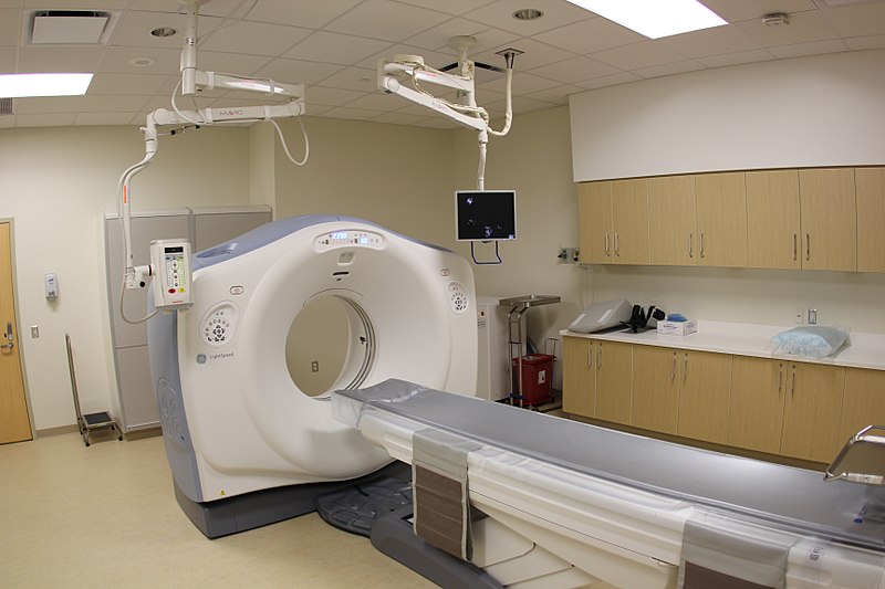
A Computed Tomography (CT) machine acquires xray images
from different angles around the head-to-toe axis of the patient.
The machine provides a stack of 2D axial xray "slices" of the
patient, giving a 3D view of the internals.
Pre-operative applications are in diagnosis, modelling, and
planning.
Intraoperative CT (iCT) can be used for tool guidance
(e.g. brachytherapy and biopsy), but out-of-plane tool tracking is
difficult as only a slice of the tool appears.
Some facts:
Slice pixels can be as small as 0.625mm x 0.625mm.
Spacing between slices should be maximized to reduce dose.
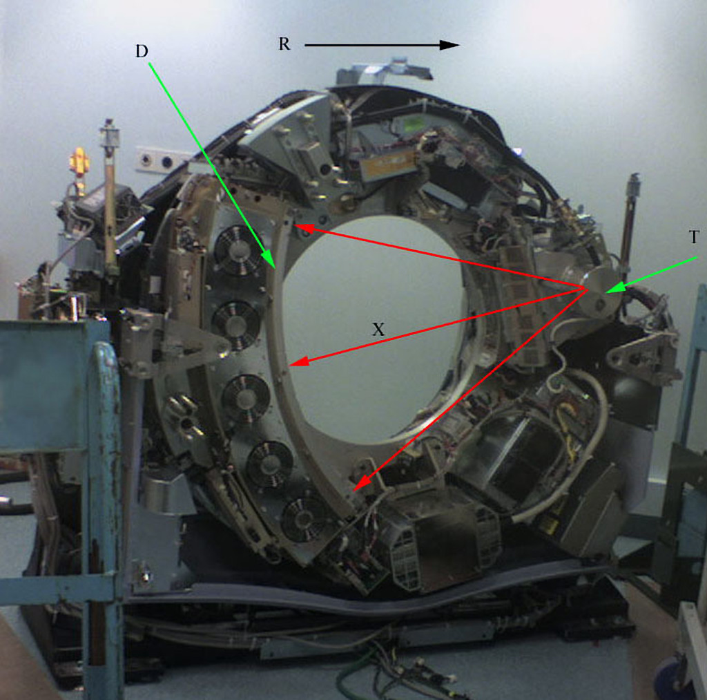
CT Machine
The CT is an xray machine that spins around the patient.
Below, $T$ is the xray tube, $X$ are the xrays, $D$ is the
detector, and $R$ is the direction of rotation.
[radiopaedia.org]
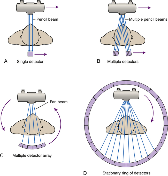
CT machines have gone through several generations of development:
[radiologykey.com]
1st: pencil beam scans linearly, then rotates and repeats.
2nd: fan beam scans linearly, then rotates and repeats.
3rd: fan beam rotates with detectors
4th: fan beam rotates, detectors fixed.
5th: all fixed sources and detectors.
6th: helical scan
Sinograms
The raw output for one CT slice is a 2D "sinogram", $g(\theta,\rho)$:
Each sinogram row corresponds to a particular angle, $\theta$, of the xray apparatus.
Each sinogram column corresponds to a position, $\rho$, on the xray detector.
Angles range in only [0,180] degrees because the rays at angles
$\theta$ and $\theta + 180$ have almost the same
attenuation. (Why not the same?)
 [wikipedia]
 [radiopaedia.org]
 [radiologykey.com]